
 MENU
MENU 

How to color-code nearly invisible nanoparticles
With a bit of metal, nanoparticles shine in colors based on size.
 Enlarge
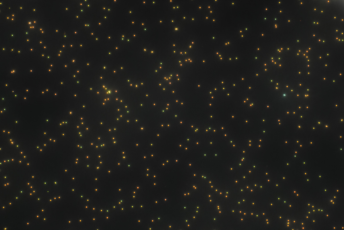
Color-coding is often used as a simple way to organize items that allow for quick understanding, like file folders, subway lines, and flavors of colorful candy. Now, researchers at the University of Michigan are applying color-coding to particles that are about the size of color itself, allowing scientists to quickly determine the size of nanoparticles. This can help in biomedical drug delivery, biological sensors, advanced coatings, and lithography of more advanced computer chips.
Nanoparticles used for these purposes, such as silica nanoparticles, typically have a low refractive index, meaning they do not interact strongly with light, because they are near the same size as the wavelengths of visible light–from 390 nanometers to 700 nanometers. This makes them difficult to discern with light.
However, the U-M research team, led by L. Jay Guo, professor of Electrical and Computer Engineering, has discovered a way to make nanoparticles shine in a range of colors based on their size. By adding metal caps to each particle, smaller nanoparticles around 421 nanometers in diameter appear green, while larger ones around 486 nanometers give off a red hue. The coloring is exact enough that researchers can tell a size difference as small 8 nanometers based on color.
Besides acting as a quick visual marker for nanoparticle size, the method could allow researchers to see color changes occur when the nanoparticles carry payloads due to a changes in their refractive index. This could help in biomedical drug delivery by making sure nanoparticles are both picking up drug molecules and dropping them off on target simply by looking at the change of their color. The same color signal could alert biological sensors to a change in state.
This work is also important in the so-called nanosphere lithography for computer chips and sensors, where nanoparticles have stringent requirements to be uniform in size in order to create the proper patterns to print circuits.
And finally, both the size of nanoparticles, and number of each size, change the properties of advanced coatings. This can make a surface smoother or rougher, repel or absorb water, or reflect or trap light.
To accomplish the color-coding, Guo and his team first had to distinguish what’s called the Mie resonances of the nanoparticles, which are the sizes that scatter light particularly strongly or, as in this case, weakly. Then, the researchers enhanced the Mie resonance to become observable as visible light.
The team began by coating a glass slide with a thin layer of gold. Then, silica nanoparticles were sprinkled on the slide, and finally, were capped with another thin layer of gold. This process increased the ability of the nanoparticles to confine light, and made them visible when viewed with a dark-field microscope (which shows only the light returning from the particles).
Currently, sizing of particles is completed through a process called dynamic light scattering, where a laser is shot at a sample and size distributions are measured over time, or with a scanning electron microscope if they are in the nanoscale. This new method is more accurate than dynamic light scattering, which can only resolve sizes to hundreds of nanometers. Compared to using a scanning electron microscope, which requires a slow, one-at-a-time particle sizing and expensive instrument, the new method allows for the sizing of multiple particles at once.
The work also raises questions about the conventional thinking on optical magnetic resonance, which relates to the magnetic dipole radiation of electromagnetic waves. Previously, high refractive index particles, which interact more with light, were needed to observe magnetic resonance.
“It was not possible to observe this in low index particles,” Guo said. “But with a strong resonance, we found even particles with a very low index can induce a strong displacement current.”
Jing Zhou, formerly a postdoctoral fellow advised by Guo and now faculty at Shanghai Institute of Technical Physics, Chinese Academy of Sciences, is first author on the paper that outlines the group’s work, “Visualizing Mie resonances in low-index dielectric nanoparticles.”
The work was supported in part by the National Science Foundation (grant number DMR 1120923), the National Key Research and Development Program of China (grant number 2017YFA0205801), the Hundred Talent Program of the Chinese Academy of Sciences, the National Natural Science Foundation of China (grant number 61605230), the Shanghai Pujiang Program (grant number 16PJ1410400).
The metal deposition was done in the Lurie Nanofabrication Facility.
Guo also holds appointments in the Macromolecular Science and Engineering Program, the Department of Mechanical Engineering and the Applied Physics Program.